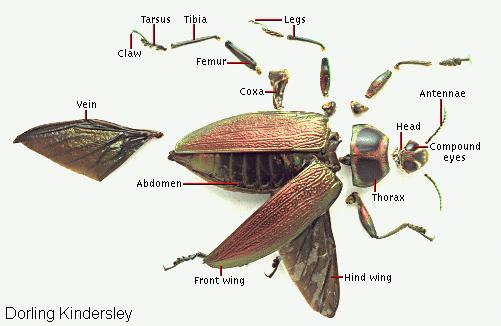

This jewel beetle has been dissected to show the various components of its anatomy. The head, or front segment, contains the mouth, eyes, and antennae. The first segment of the thorax, located just behind the head, bears the first pair of legs. The large posterior section of the body, including the second and third segments of the thorax and the abdomen, contains the remainder of the walking legs and all the vital body organs. The wings lack muscles and are manipulated by muscles located inside the abdomen. The outer surface of the body, called the exoskeleton, is protected by a hard chitinous material.

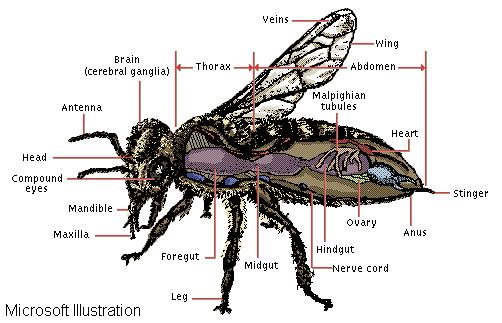
This diagram illustrates many of the important internal and external organs common to insects. Crucial elements in the insect's nervous, reproductive, circulatory, and digestive systems are labeled.

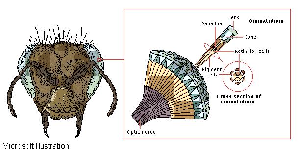
The eyes of insects and many other arthropods are compound, each composed of up to several thousand individual visual organs called ommatidia. The surface of each ommatidium is a hexagonal lens, below which is a second, conical lens. Light entering the ommatidium is focused by these lenses down a central structure called the rhabdom, where an inverted image forms on light-sensitive retinular cells. Pigment cells surrounding the rhabdom keep light from other ommatidia from entering. Optic nerve fibers transmit information from each rhabdom separately to the brain, where it is combined to form a single image of the outside world.
